Platforms for live, high-resolution imaging of organotypic tissue slices
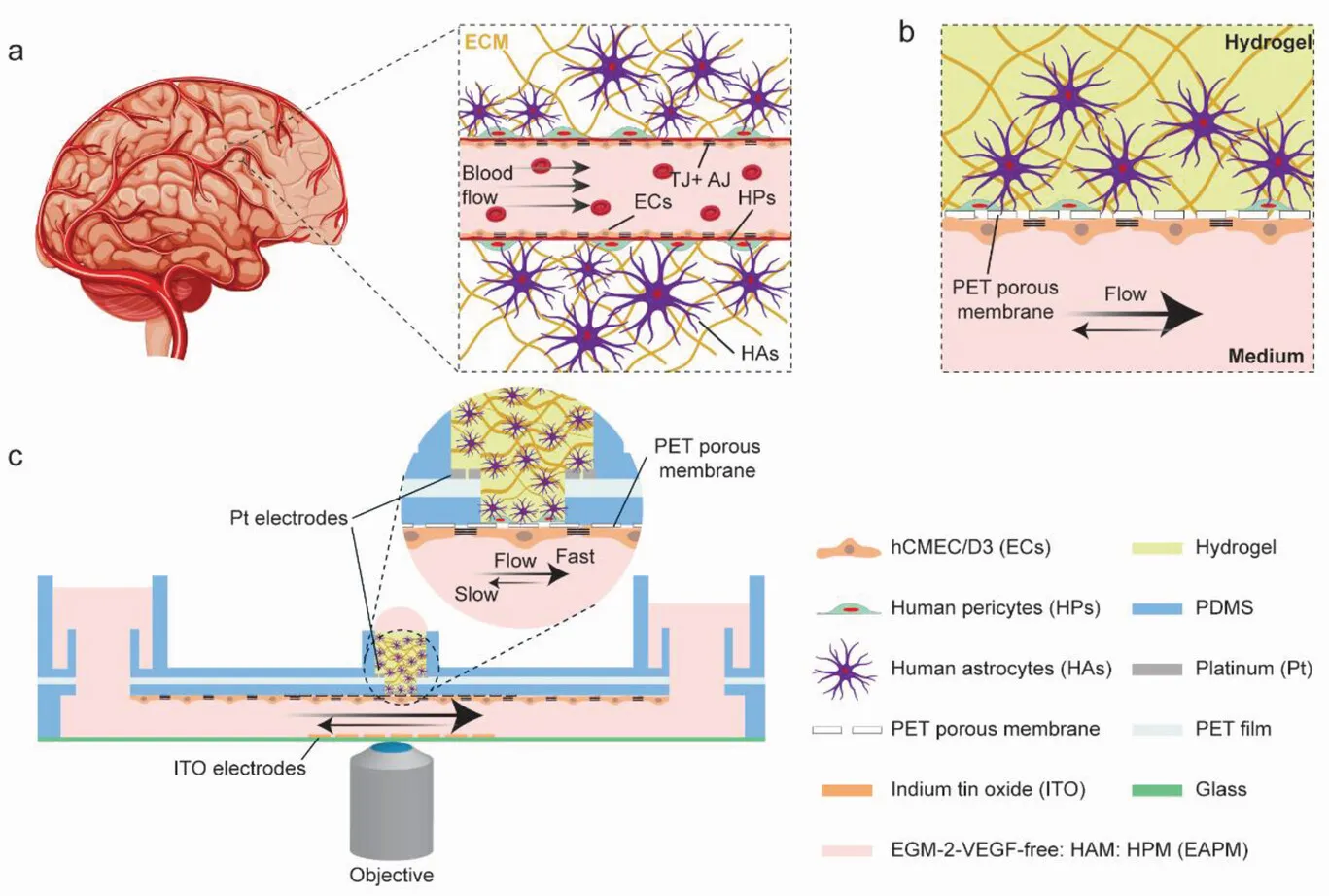
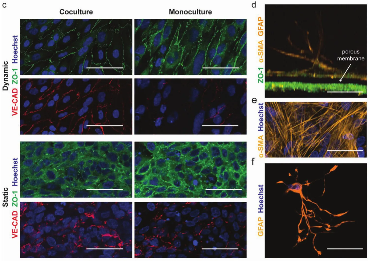
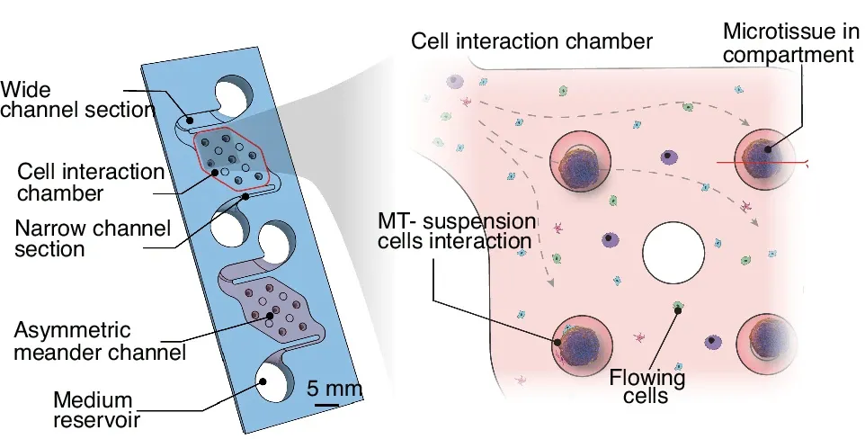
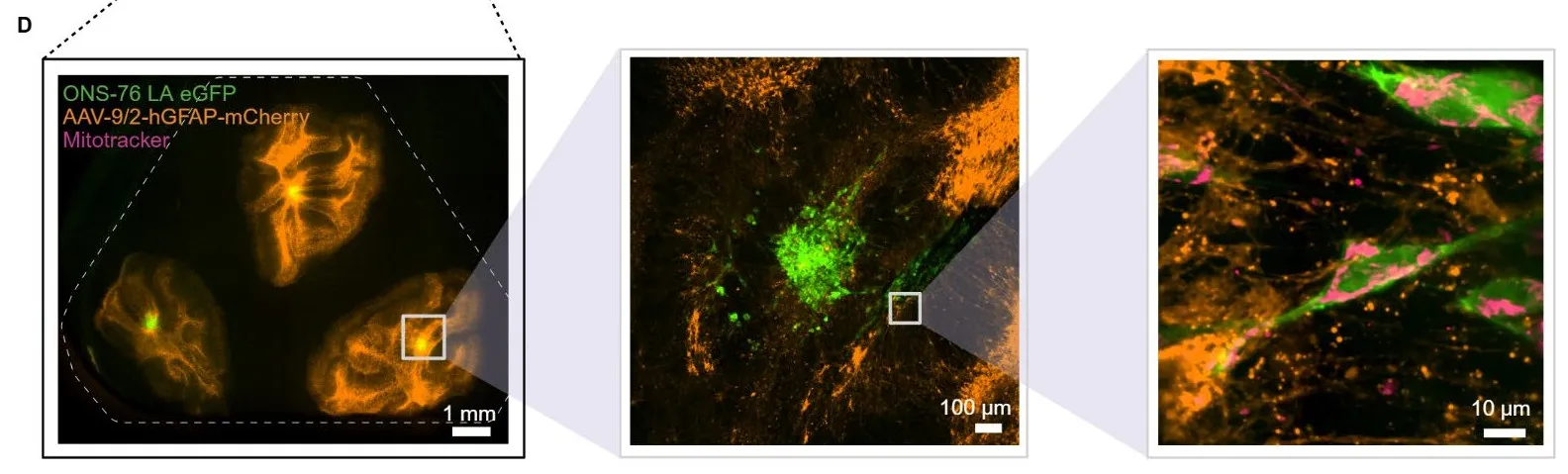
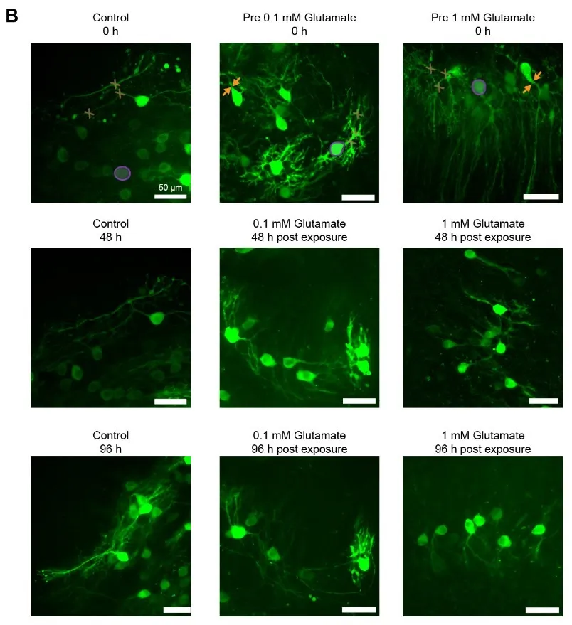
Two platforms for imaging brain tissue slices over several days. PHIROS keeps slices alive under continuous, oxygenated perfusion in a sealed chip for multiple days, enabling subcellular-resolution imaging and compound delivery with precise temporal resolution. The platform was used to visualize astrocyte calcium activity, medulloblastoma–astrocyte interactions and mitochondrial transfer between tumor cells. A newer modular insert works in standard well plates and in an automated microfluidic instrument, so the same slice can be imaged repeatedly, as demonstrated with cerebellar astrocytes and Purkinje cells exposed to glutamate. The projects were carried out in collaboration with researchers at Kispi Zurich and Lunaphore SA.
Papers: PHIROS, bioRxiv 2026 · On-demand imaging, bioRxiv 2026